Diagnosing cerebral venous thrombosis: could CT help?
Cerebral venous thrombosis (CVT) is an uncommon cause of stroke where a blood clot forms
in the veins that drain blood from the brain. Patients with CVT present in a myriad of ways, including but not limited to headaches, seizures, weakness and sensory loss.
CVT is
uncommon and difficult to diagnose, requiring complex and expensive imaging
techniques such as MRI and advanced CT imaging. However, these tests are often
not ordered in the emergency department, especially for patients with seizures
or headache, for which a multitude of other conditions could be the cause.
Researchers at the Florey are searching for better
ways to diagnose cerebral venous thrombosis. Their recently published study looks at whether plain CT is
able to detect CVT. Plain CT, unlike MRI, is fast and readily accessible in any major hospital.
 |
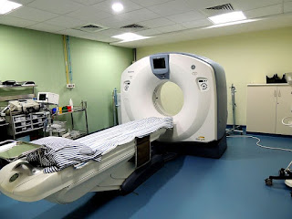
| Could fast, accessible CT scanners like this one replace MRI and advanced CT for detecting cerebral venous thrombosis? Source: Wikimedia Commons |
Unfortunately, the study found that this simpler imaging technique
wouldn’t be an effective substitute for current methods. ‘Plain CT, even
analysed using sophisticated quantitative methods, is insufficient to rule out
a cerebral venous thrombosis, a cause of stroke, headache and seizures,’
said Professor Vincent Thijs, who led the study. ‘Whenever CVT is suspected,
one should not rely on CT only.’
So the search continues for improved ways of diagnosing CVT.
‘Better screening techniques for this condition could include blood biomarkers
in combination with imaging findings,’ said Professor Thijs.
Read the full study here.
Comments
Post a Comment